Labeling the molecules that modify the electrode surface with a fluorophore enables study of this adsorbed layer using fluorescence. Moreover, the study of the spatial distribution, or heterogeneity of the surface coverage is also possible.
A schematic of the setup is shown:
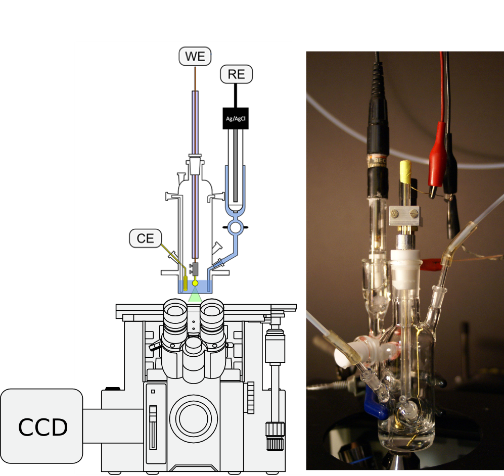
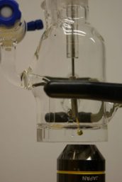
The spectroelectrochemical cell is specially made by our glassblower Brian Ditchburn. He is able to put a very thin (0.25 mm) cover glass like window onto the bottom of the cell which enables high quality optical resolution images.
An overview of this approach with more technical details is provided in:
-
Casanova-Moreno, J.; Yu, Z. L.; Massey-Allard, J.; Ditchburn, B.; Young, J. F.; Bizzotto, D. Luminescence in Electrochemistry, Applications in Analytical Chemistry, Physics and Biology. Luminescence in Electrochemistry 2017, 7(2), 21–77. https://doi.org/10.1007/978-3-319-49137-0_2.
- Bizzotto, D. In Situ Spectroelectrochemical Fluorescence Microscopy for Studying Electrodes Modified by Molecular Adsorbates. Current Opinion in Electrochemistry 2018, 7, 161–171. Link to article
- Bizzotto, D.; Yang, Y.; Shepherd, J. L.; Stoodley, R.; Agak, J. O.; Stauffer, V.; Lathuilliere, M.; Akhtar, A. S.; Chung, E. J. Electroanal. Chem. 2004, 574, 167.
- Bizzotto, D.; Shepherd, J. L. Advances in Electrochemical Science and Engineering, 2006; Vol. 9, pp. 97–126. DOI
We have used this method extensively. See the following pages for more information