Self Assembled Monolayers (SAMs) adsorbed onto gold electrode surfaces has been studied quite a bit with electrochemical methods, and even using spectroelectrochemistry, but not often with spatial resolution less than 0.1mm. Using fluorescence microscopy and our in-situ methods (more here) we showed that the SAM can be removed from various regions of the electrode in a potential specific manner.
- Shepherd JL, Kell A, Chung E, Sinclar CW, Workentin MS, Bizzotto D. Selective reductive desorption of a SAM-coated gold electrode revealed using fluorescence microscopy. J Am Chem Soc. 2004;126(26):8329–8335. doi:10.1021/ja0494095.
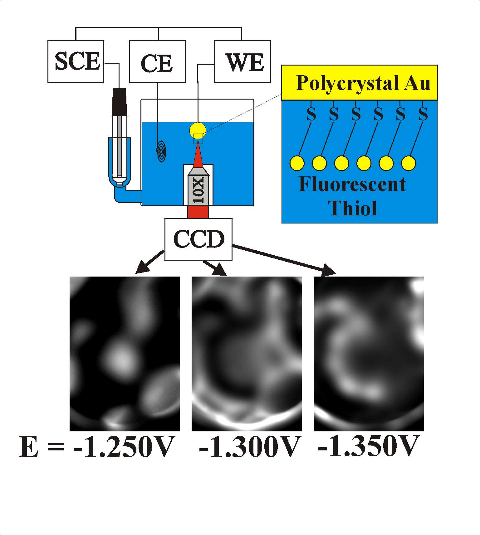
Reductive desorption from amulticrystalline Au bead electrode. Notice how the fluorescence images at more negative potentials are mutally exclusive.
This method was also applied to the oxidative desorption of the same thiol SAM
- Musgrove A, Kell A, Bizzotto D. Fluorescence imaging of the oxidative desorption of a BODIPY-alkyl-thiol monolayer coated Au bead. Langmuir. 2008;24(15):7881–7888. doi:10.1021/la800233c.
Recently, we were able to do the same measurements on a single crystal Au bead electrode. This spherical surface displayed surface features that were consistent with the whole stereographic triangle. We were able to map the surface and correlate the desorption potential to the surface structure. Using three different types of SAM forming adsorbates, the extent of intermolecular interaction and the influence of the surface on the SAM formation was discussed.
- Yu ZL, Casanova-Moreno J, Guryanov I, Maran F, Bizzotto D. Influence of surface structure on single or mixed component SAMs via in-situ spectroelectrochemical fluorescence imaging of the complete stereographic triangle on a single crystal Au bead electrode. J Am Chem Soc. 2014. doi:10.1021/ja5104475.
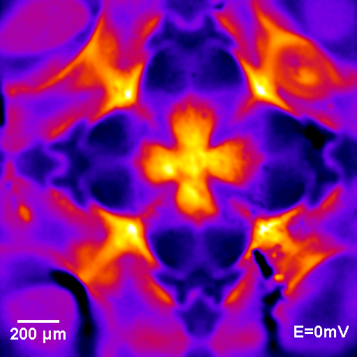
MCH/ssDNA mixed component SAM on a single crystal Au bead electrode. The symmetry of the image is a sign of the single crystal nature of the surface. The 100 surface is in the centre, the 111 facets are at the four corners, with the other surfaces related to these by symmetry